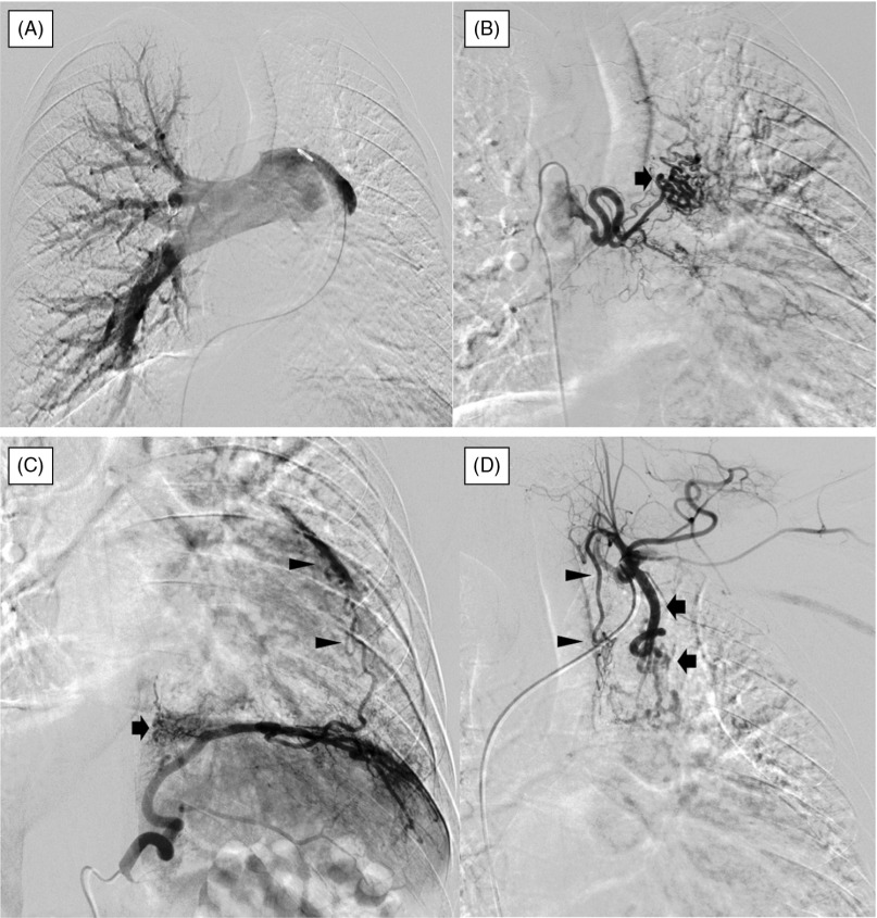
Fig. 3.
Angiography. (A) Angiography of the pulmonary artery: Although the right pulmonary artery is depicted, the left pulmonary artery is completely absent from its origin. (B) Angiography of the left bronchial artery: The artery originates from the descending thoracic aorta at the level of the tracheal bifurcation and is highly dilated. Blood vessels to the left superior lobe were dilated and tortuous (arrow). (C) Angiography of the left inferior phrenic artery: The inferior phrenic artery is highly dilated and collateral vessels ascending along the left pulmonary basilar region (arrow) and left cardiac margin (arrowheads) are depicted. (D) Angiography of the left subclavian artery: The left internal thoracic artery is highly dilated and tortuous (arrows). The vessels that originate from the thyrocervical trunk and are distributed to the left aspect of the trachea are dilated (arrowheads).

