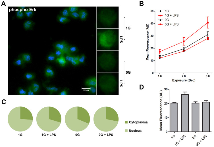
Figure 4. Effects of microgravity on ERK activation.
The figure shows phospho-Erk analysis of LPS stimulated monocytes in the presence (1 G) or relative absence of gravity (0 G). Fixed monocytes were stained fluorescently (green) and the nuclei were visualized by DAPI (blue) (Figure 4A). Comparison between individual cells clearly shows a brighter staining when stimulated with LPS in the presence of gravity. Staining intensity was measured at different exposure times (Figure 4B) and mean fluorescence was finally determined at 2000 ms (Figure 4D). Microgravity impairs the activation of Erk in monocytes. Analysis of nuclear localization of activated Erk shows no change (Figure 4C). The graphs are based on 32 determinations.

