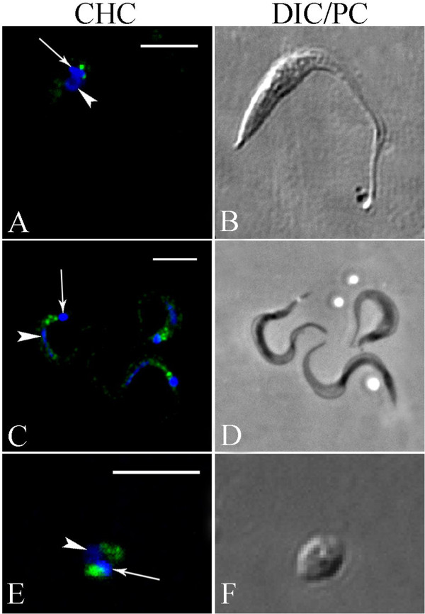
Figure 2.

Analysis of TcCHC localization in Trypanosoma cruzi using a TcCHC monoclonal antibody. Subcellular localization of TcCHC in the various developmental forms of T. cruzi. (A-B): epimastigote. (C-D): metacyclic trypomastigotes. (E-F): isolated amastigote. Note strong immunolabeling in a region adjacent to the kinetoplast (arrow), where the flagellar pocket and Golgi complex are located. Nucleus (arrowhead) and kinetoplast DNA were stained with Hoechst 33342. A: rabbit anti-mouse IgG conjugated to Alexa Fluor 594 (pseudocolored in green); C &E: rabbit anti-mouse IgG conjugated to Alexa Fluor 488; B &F: Differential interference contrast (DIC) images of the parasite body; D: phase contrast images of the parasite body. Scale bars = 5 μm.
