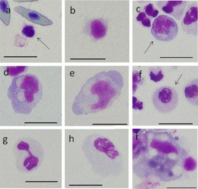
Fig. 3.

Cells from peripheral blood (a-h) and spleen (i) of ayu, Plecoglossus altivelis altivelis, after Leishman-Giemsa staining. Bar=10 µm, a: A1 type (arrow), b: A2 type, c: B1 type (arrow), d: B2 type, e: B3 type, f: C1 type (arrow), g: C2 type, h: C3 type and i: F type.
