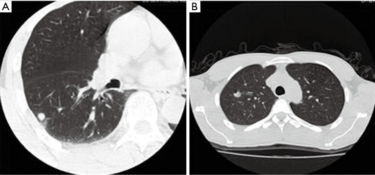
Figure 1.

<10 mm lung nodules: (A) right inferior pulmonary nodules of a 57-year-old man, axial CT image showed: smooth nodule edge, no lobulation or glitches, local pleural stretch, and adjacent few fiber lesions. The prediction P value was 0.312, and lesion was considered to be a benign pulmonary nodule. It was later identified to be a pulmonary tuberculosis mass in the right inferior lung, as demonstrated by pathological examinations; (B) left upper lung nodule in a 37-year-old man, axial CT image showed: The nodule was lobular, with air cavity density and adjacent vascular aggregation inside it. The prediction P value was 0.647, and lesion was considered to be a malignant pulmonary nodule. It was pathologically confirmed to be the organizing pneumonia in the right upper lung. CT, computed tomography.
