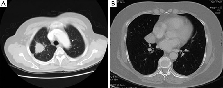
Figure 3.

>20 mm lung nodules: (A) right upper pulmonary nodules in a 61-year-old man. Axial CT image showed: the nodule was lobular, along with glitches at its edge, pleural indentation, and vascular aggregation. The prediction P value was 0.930, and the lesion was considered to be a malignant pulmonary nodule. It was pathologically confirmed to be a poorly-differentiated adenocarcinoma in the right upper lung; (B) right middle pulmonary nodules in a 66-year-old man. Axial CT image showed: smooth nodule edge, but without lobulation, glitches, vascular aggregation, or pleural indentation. The prediction P value was 0.297, and lesion was considered to be a benign pulmonary nodule. The lesion was pathologically confirmed to be a spindle cells carcinoid in the right middle lung. CT, computed tomography.
