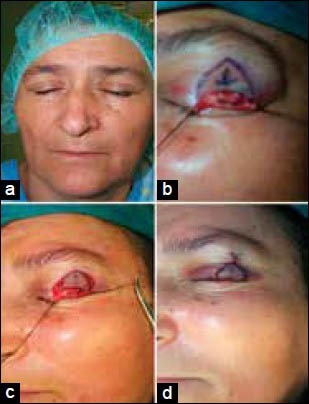
Figure 1.

(a) Preoperative appearance of the tumoral lesion in the left upper eyelid, (b) Appearance of the planning after excision, (c) Appearance of the split thickness upper eyelid defect after excision and harvested orbicularis oculi myocutaneous V-Y advancement flap, (d) Postoperative appearance of the normal closure of the eyelid
