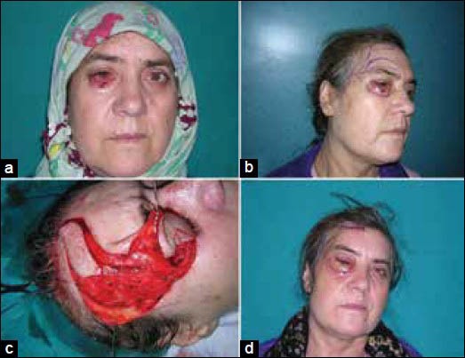
Figure 3.

(a) Preoperative appearance of the tumoral lesion in the right lower eyelid, (b) Appearance of the preoperative two-different planing, (c) Appearance of the full thickness lower eyelid defect, after excision, and harvested superficial temporal artery frontal branch based island flap, (d) Postoperative appearance of the early result
