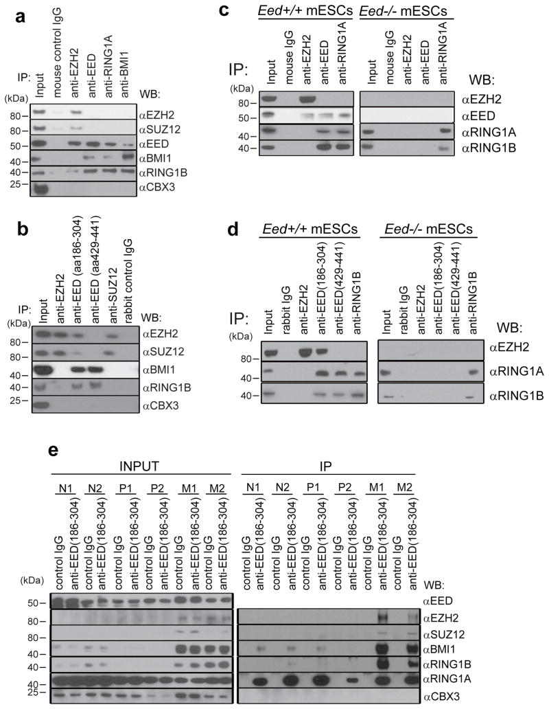
Figure 1. PRC2 protein EED interacts with PRC1 proteins.
a, Immunoprecipitation of DU145 cell lysates with the indicated mouse monoclonal antibodies anti-EZH2, anti-EED (Millipore, cat# 05-1320), anti-SUZ12, anti-BMI1, anti-RING1A and control IgG was followed by immunoblot analysis. Anti-CBX3 was used as a negative control. Representative experiment from at least three independent analyses is shown for this and subsequent analyses. b, Immunoprecipitation of DU145 lysates with the indicated rabbit polyclonal antibodies anti-EZH2, anti-EED (aa186-304) (Santa Cruz, cat# sc-28701), anti-EED (aa429-441) (Millipore, cat# 09-774), anti-SUZ12 and control IgG was followed by immunoblot analysis. c, Immunoprecipitation of Eed+/+ and Eed−/− mESCs cell lysates with the indicated mouse monoclonal antibodies anti-EZH2, anti-EED (Millipore, cat# 05-1320), anti-RING1A and control IgG was followed by immunoblot analysis. Representative experiment from at least three independent analyses is shown for this and subsequent analyses. d, Immunoprecipitation of Eed+/+ and Eed−/− mESCs cell lysates with the indicated rabbit polyclonal antibodies anti-EZH2, anti-EED (aa186-304) (Santa Cruz, cat# sc-28701), anti-EED (aa429-441) (Millipore, cat# 09-774), anti-RING1B and control IgG was followed by immunoblot analysis. e, Immublot analysis of endogenous EED pull down (rabbit control IgG or anti-EED aa186-304) using lysates from normal prostate (N1 and N2), localized prostate cancer (P1 and P2) and metastatic prostate cancer tissues (M1 and M2).

