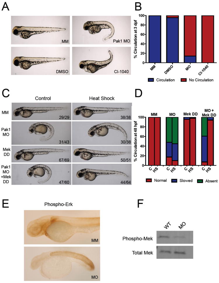
Figure 3. Pak1 signals through the Erk pathway in heart development.

(A) Comparison of chemical inhibition of Mek and pak1 morphants at 3 dpf. WT embryos were placed in egg water containing DMSO or 1 μM CI-1040 at the 1-cell stage. The water was changed every 24 hours with new drug. The embryos were analyzed for gross morphology and presence or absence of circulation. (B) Quantification of circulation seen with Mek inhibition at 48 hpf. (C) Representative images of 48 hpf pak1 morphants rescued by an active form of Mek (Mek DD). An inducible Mek1 DD expression plasmid was coinjected with the pak1 MO at the one-cell stage, followed by heat-shock at 24 hpf as indicated. (D) Immunohistochemistry for phosphorylated Erk.
