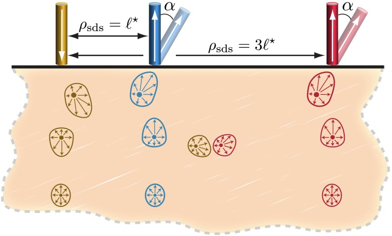
Fig. 3.
Illustration of the model tissue with a single source and two detectors at different distances and two detection angles. Additionally, characteristic forward and adjoint radiance cross sections are displayed for the source and normally oriented detectors. The forward and adjoint radiances display the high degree of direction anisotropy in locations near the source/detector.

