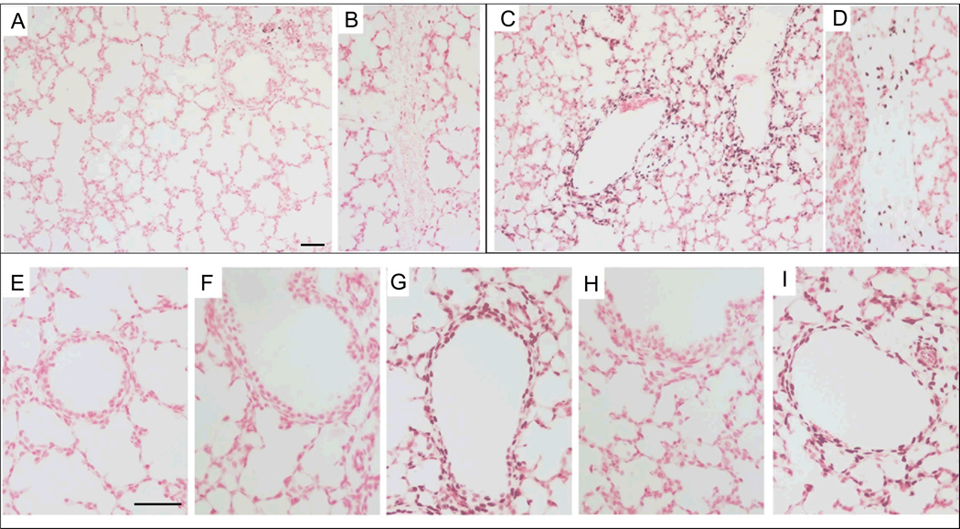
Figure 2. Egr-1 protein increased around bronchioles with mechanical ventilation.
(A–B, E) Control lambs had only occasional Egr-1 staining in the (A) vessels and none was found in (B) intra-lobar connective tissue (20×). (C–D) Egr-1 signal increased with mechanical ventilation with nitrogen (N2) and was localized to cells around bronchioles (C) and in cells within connective tissue (D) (20×). There was minimal staining in the distal epithelium. (E–I) Higher power images of Egr-1 staining. Lambs receiving VT ventilation with N2 (G) and O2 (I) demonstrate increased Egr-1 surrounding small airways. CPAP exposure, with N2 (F) or O2 (H) did not increase staining. Scale bar 50 µm.

