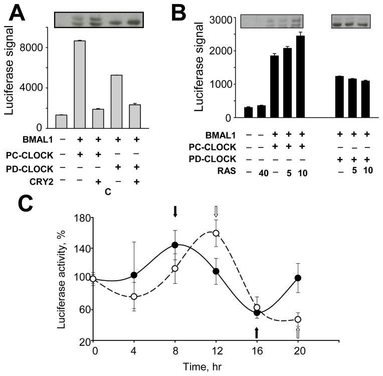
Fig. 5. Stabilization of phospho-deficient CLOCK affects CLOCK/BMAL1 functional activity.
A. Phospho-deficient CLOCK proteins are less transcriptionally active compare to phospho-competent CLOCK. 293T were transfected with a GAL4 luciferase reporter and plasmid encoding BMAL1 with either GAL4-PC-CLOCK fusion or GAL4-PD-CLOCK fusion S427/429/431/433/434/436A, followed by luciferase activity measurement. Bars represent relative luciferase signal normalized for efficiency of transfection using β-Gal assay. Experiments were performed at least 3 times in duplicates. Values are mean ± standard error. The transactivation property of all phospho-deficient mutants tested was significantly decreased compared to wild-type CLOCK (p<0.05 as determined by Student’s t-test). The insert shows the abundance and pattern of phosphorylation of CLOCK proteins analyzed by Western Blot.
B. Over-expression of RAS increases transcriptional activity of phospho-competent but not phospho-deficient CLOCK proteins.
A similar GAL4 luciferase assay was executed with co-expression of BMAL1 and either GAL4-CLOCK fusion or GAL4-PD-CLOCK fusion S427/429/431/433/434/436A. Increasing concentrations of the plasmid expressing RAS were used as indicated. Transfected cells were lysed in reporter buffer and luciferase activity was measured and normalized for β-Gal activity. The Western Blot inserts show the abundance and phosphorylation status of CLOCK.
C. Stabilization of phospho-deficient CLOCK protein delays the phase of oscillation in synchronized fibroblasts.
MEFs derived from mice with the targeted disruption of the Clock gene were transfected with mPer1-luciferase reporter gene and BMAL expression construct with either wild type or PD-CLOCK mutant S434/436/437/440/441A. 36 hrs post transfection cells were transferred to 24-well plates and treated with 0.1 uM dexamethasone for 4 hrs. Luciferase activity was measured in cell lysates over a 20-hr period with a 4-hr resolution. Shown is a representative experiment out of three that were each performed in triplicates. Values are normalized by luciferase signal in cells at 0 time point (time of dexamethasone removal) and show mean value ± standard error. Closed circles represent cells transfected with PC-CLOCK, open circles – cells transfected with PD-CLOCK. Black and white arrows indicate peak and trough of Per1-driven luciferase expression by the phospho-competent and phospho-deficient CLOCK/BMAL1 complexes respectively.

