
Extended Data Figure 6. The PKD1-L1/PKD2-L1 channel is highly temperature sensitive.
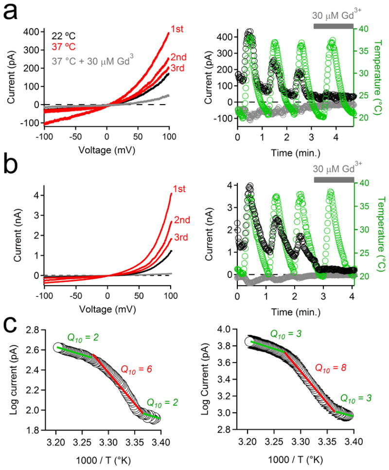
(a, b) The effects of repeated temperature stimulations from 22 to 37°C on the PKD1-L1/PKD2-L1 current recorded from (a) RPE primary cilia and (b) HEK-293T cells (plasma membrane) transfected with PKD1-L1 and PKD2-L1. Left, Currents elicited by a series of 1 Hz voltage ramps from -100 to +100 mV from 21 to 38°C in control conditions (red traces) or in the presence of 30 μM Gd3+ (grey trace). Right, resulting current amplitudes (-100 mV, grey circles; +100 mV, black circles) and cilia temperature (green circles) are plotted as a function of time. Grey bar indicates the duration of extracellular 30 μM Gd3+ application. (c) Arrhenius plots of the PKD1-L1/PKD2-L1 currents recorded from (left) RPE primary cilia and (right) when heterologously expressed in HEK-293T cells. Q10 values were derived from 3 linear fits of the average normalized current magnitude from 3 phases of the thermal response (21-24°C; 24-32°C; 32-38°C; ± SEM; n = 4 cilia or 4 cells).