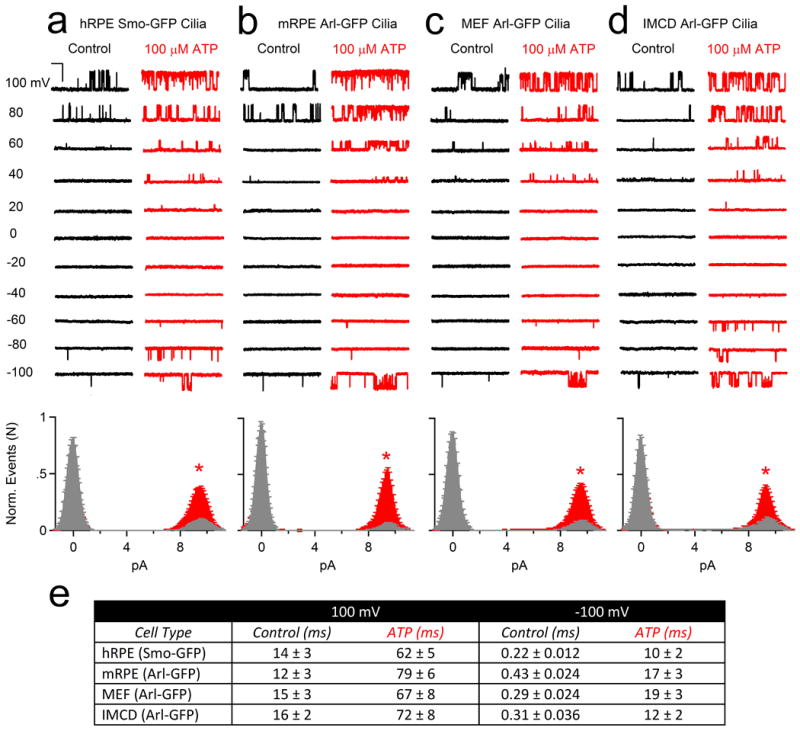
Extended Data Figure 2. ATP indirectly activates the cilia conductance from four different cell types.

Top, Single channel currents activated by 1.5 s depolarizations to the indicated potentials in control (black traces) and 100 μM extracellular ATP (red traces) recorded from primary cilia derived from (a) human RPE Smo-GFP cell lines, (b) mouse RPE Arl-GFP primary cells, (c) mouse MEF Arl-GFP primary cells, (d) mouse kidney IMCD Arl-EGFP cells (scale = 10 pA and 200 ms). Bottom, corresponding open probability histograms measured in control (grey) and in the presence of 100 μM ATP (red; ± SEM, n = 4-6 cilia, asterisks indicates P < 0.005). (e) Average open dwell times measured from the cilia of these four cell types in control and ATP conditions.
