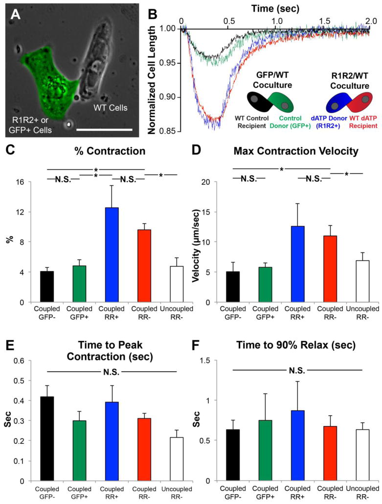
Figure 5. R1R2-hESC-CMs enhance contractility of neighboring WT cardiomyocytes.
HESC-CMs were transduced with R1R2-GFP or control GFP adenovirus and cocultured with WT hESC-CMs. (A) The contractile parameters of each cell in a heterogeneous GFP+/GFP− doublet were measured. As a control, measurements were also taken from a WT uncoupled cell in the R1R2 cultures (white bar). (B) Representative traces show that R1R2-GFP+ cells and coupled WT hESC-CMs exhibit increased (C) magnitudes and (D) velocities of contraction but no change in the kinetics of (E) contraction or (F) relaxation. n=3–12 per condition. *p<0.05 N.S. not significant

