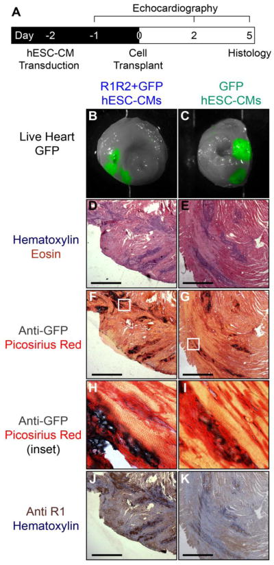
Figure 7. In vivo transplantation of R1R2-hESC-CMs.
(A) hESC-CMs were transduced with R1R2-GFP or GFP on D-2 and 15e6 cells transplanted into healthy nude rat hearts on D0. Animals were monitored echocardiographically before and after cell transplantation, and on D5 the hearts were explanted and histology performed. (B,C) 100% of animals receiving grafts had positive GFP signal upon gross sectioning and imaging. (D,E) H&E demonstrated areas of dense nuclei staining consistent with engrafted cells. (F,G) Anti-GFP staining colocalized to the same graft regions identified by H&E. (H,I) High magnification inset of these stains show clear regions of GFP+ signal directly adjacent to host myocardium. (J,K) Staining for the R1 subunit in serial sections revealed strongly positive clusters of cells in R1R2-GFP animals, but only weak signal in GFP-hESC-CM control hearts. Scale bar 500 μm

