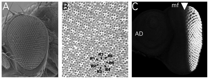
Figure 1. Summary of Drosophila eye structure and development.

A). Scanning Electron Micrograph of the adult eye, with anterior to the left. Each adult compound eye contains ~750 facets, or ommatidia.
B). Thick section through an eye revealing the repetitive cellular pattern. The rhabdomeres of seven of the eight photoreceptor neurons are apparent in each plane of section, and are labeled for one ommatidium.
C). Eye-antennal imaginal disc from a mid-third instar larvae (~96h after egg laying). Clusters of differnetiating photoreceptor neurons behing the morphogenetic furrow (arrowhead) are labeled with antibody against ELAV. The morphogenetic furrow moves from posterior to anterior (left ot right in this image) across the eye disc. Ahead of the morphogenetic furrow, individual cell fates remain unspecified and no ELAV labeling is seen. The antennal disc (AD) is also unlabeled.
