Figure 3.
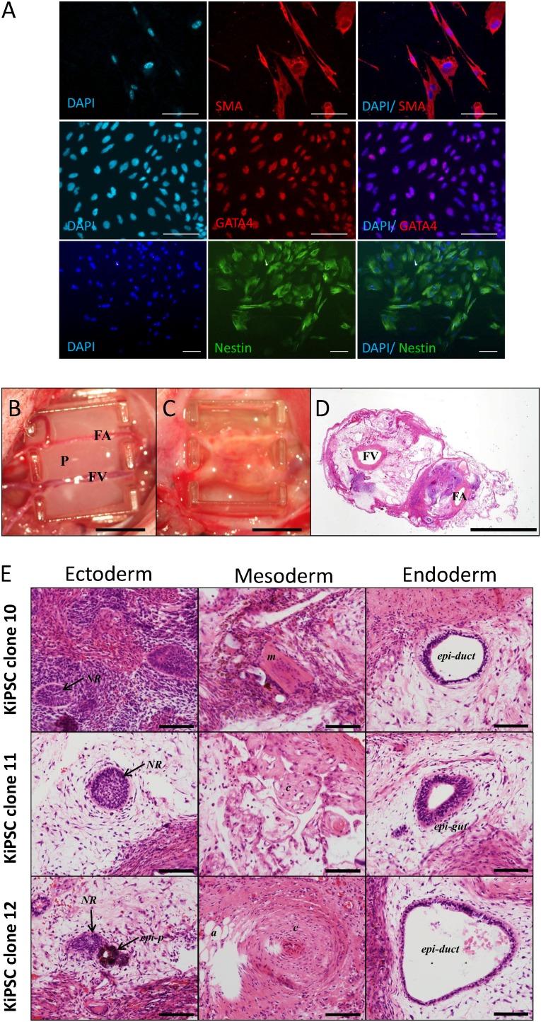
KiPSC clones retain in vitro and in vivo differentiation potentials. (A): Immunocytochemistry analysis of in vitro differentiation potential of established KiPSCs into the three germ layers: endodermal (GATA4), mesodermal (SMA), and ectodermal (Nestin) lineages. Scale bar = 100 μm. (B, C): Image of the tissue engineering chamber implanted with KiPSCs (B) and teratoma formation after 4 weeks (C). Scale bar = 5 cm. (D): Hematoxylin and eosin staining of teratoma constructs harvested at 4 weeks after implantation. Scale bar = 2 mm. (E): In vivo teratoma formation of established KiPSCs showing cells representative of the three germ layers. Scale bar = 100 μm. Abbreviations: a, adipose; c, cartilaginous structure; DAPI, 4′,6-diamidino-2-phenylindole; epi-duct, epithelial-lined duct structure; epi-gut, gut-like epithelium; epi-p, pigment epithelium; FA, femoral artery; FV, femoral vein; KiPSC, hiPSC colonies generated from keratinocytes; m, muscle; NR, neural rosette structure; P, rat plasma clot containing cells.

