Figure 1.
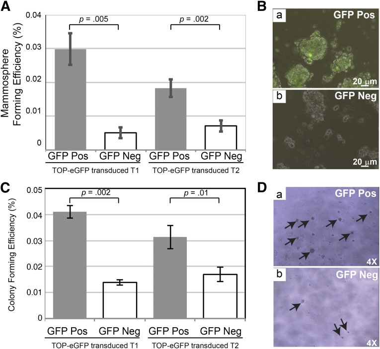
Mammosphere and Matrigel assays of sorted subpopulations of TOP-eGFP lentivirus-transduced p53-null mammary tumors. (A): Sorted eGFP-positive and -negative cells were plated and grown for 7 days before they were trypsinized and replated at 5,000 cells per well into six-well ultralow attachment plates. Secondary mammospheres were counted on days 7 and 8 from each subpopulation. There are three biological and three technical replicates from each tumor. Left: T1 tumor, p = .005. Right: T2 tumor, p = .002. (B): Pictures were taken on day 8 after plating secondary mammospheres from the TOP-eGFP-positive and -negative cells from TOP-eGFP transduced T2 tumor. Scale bars = 20 μm. (C): Sorted eGFP-positive and -negative cells were plated and grown on growth factor-reduced Matrigel for 6 days at 2,500 cells per well in 96-well plates. There were three biological and three technical replicates from each tumor. Left: T1 tumor, p = .002. Right: T2 tumor, p = .01. (D): Pictures were taken on day 4 after plating the TOP-eGFP-positive and -negative cells from TOP-eGFP transduced T2 tumor. The p values were obtained by paired sample t test. Abbreviations: eGFP, enhanced green fluorescent protein; GFP, green fluorescent protein; Neg, negative; Pos, positive.

