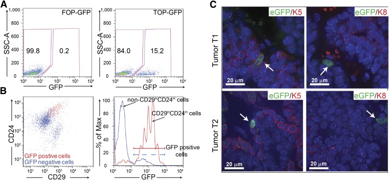
Figure 2.
Characterization of the Wnt-responsive cells in the p53-null mammary tumors. (A): Of tumor cells, 11%–16% show GFP expression following transduction with lentiviral TOP-eGFP as compared with little to no expression in the control lentiviral FOP-eGFP-transduced T2 tumor. (B): Fluorescence-activated cell sorting analysis showed that the majority (60%–80%) of eGFP-positive cells were CD29HCD24H (left) and most CD29HCD24Hcells were also eGFP-positive (right) in T2 tumor. (C): Anti-K5 (left) and anti-K8 (right) staining of the TOP-eGFP transduced tumors, T1 and T2. Scale bars = 20 μm. Abbreviations: eGFP, enhanced green fluorescent protein; GFP, green fluorescent protein.

