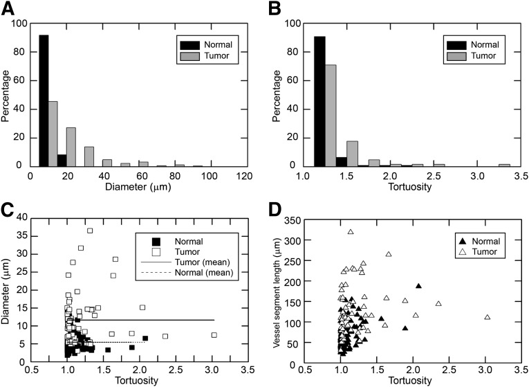
Figure 4.
Morphological characterization of vessel segments in normal mammary tissues and in p53-null tumors. (A): Distributions of vessel diameters in normal mammary glands and T1 tumors. The distribution of vessel diameters in tumors is broad as opposed to the distribution of diameters in normal mammary tissues. The two distributions are significantly different (p < .0001, chi-square test). (B): Distributions of tortuosities of vessel segments in normal mammary tissues and in T1 tumors. The two distributions are not significantly different at α = 0.05 (p = .07, chi-square test). (C): Scatter plot of diameters of vessel segments as a function of their tortuosities. (D): Plot of arc lengths of vessel segments as function of their tortuosities.

