Figure 5.
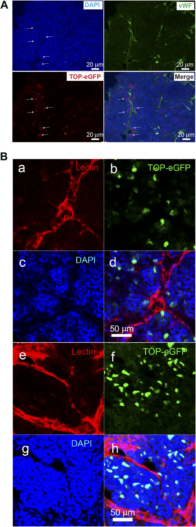
Localization of Wnt-responsive cells with respect to vessels in p53-null tumors. (A): Costaining of the vWF and anti-green fluorescent protein on the p53-null T1 tumor. Scale bars = 20 μm. (Ba–Bd): Confocal images of a region in a T1 tumor. The vessels are labeled with tomato lectin (Ba), the Wnt reporter TOP-eGFP labels the cancer stem cells (Bb), the nuclei are labeled with DAPI (Bc), and merge of the vessels, Wnt reporter TOP-eGFP labeled cancer stem cells and nuclei (Bd). (Be–Bh): Confocal images of a region in a T2 tumor. The vessels, CSCs, and nuclei are labeled with lectin (Be), TOP-eGFP (Bf), DAPI (Bg), and merge of lectin, TOP-eGFP and DAPI (Bh). respectively. Abbreviations: DAPI, 4′,6-diamidino-2-phenylindole; eGFP, enhanced green fluorescent protein; vWF, von Willebrand factor.

