
Fig. 12.
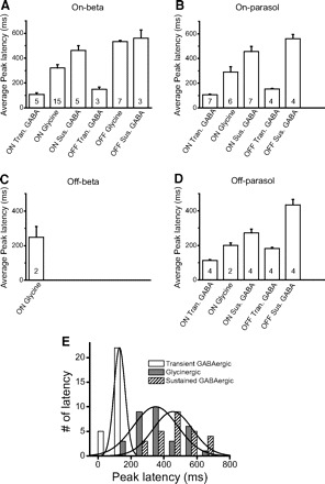
A–D: average peak latencies of different inhibitory currents. In the off beta cell, we only show the latency from on glycinergic currents because the weak GABAergic currents are hard to measure. The numbers in each bar indicate how many cases we average in that condition. Tran., transient; Sus., sustained. E: the distributions of peak latencies of transient GABAergic (open), glycinergic (gray) and sustained GABAergic (hatched) inhibition. Bin size, 100 ms. Three solid lines are the curve fitting of normal distribution.