
Fig. 1.
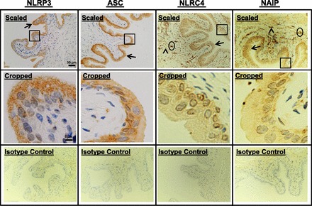
Detection of inflammasomes in the bladder by immunocytochemistry. Bladders from untreated rats were fixed, sectioned (5 μm), stained with the indicated antibodies, and counterstained with hematoxylin. Digital micrographs (2,560 × 1,920 pixels) were then taken at ×20 magnification. Scaled images (top) represent the entire micrograph scaled to 9.3% of the original size. Arrows indicate staining in urothelia, arrowheads indicate staining in the detrusor, and oval regions represent areas of staining in vascular elements. Areas encompassed by rectangles were cropped out of the full-sized micrographs and presented full scale in cropped images (middle). Isotype control images (bottom) were stained with the respective isotype antibodies as described in materials and methods. Micrographs were reduced to 9.3% of the original size to accurately serve as controls for the scaled images. All micrographs are from a representative experiment repeated three independent times. NLR, Nod-like receptor; ASC, apoptosis-associated speck-like protein containing a COOH-terminal caspase recruitment domain; NAIP, NLR family apoptosis inhibitory protein.