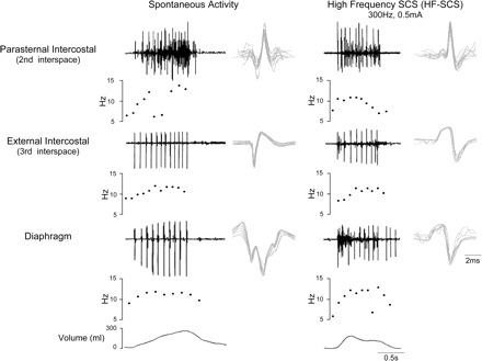
Fig. 5.

SMU activity recorded from the parasternal intercostal muscle, external intercostal muscle, and diaphragm during spontaneous breathing (left) and during HF-SCS (right) in one animal. EMG, instantaneous motor unit discharge frequency, and volume are plotted. See text for further explanation.
