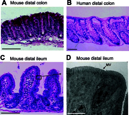
Fig. 3.

Histological evaluation of intestinal specimens after 1 h incubation in the perfusion chamber. A–C: hematoxylin/eosin staining of mouse colon, human colon, and mouse ileum. In mouse ileum, the black box represents the approximate location of the image in D. D: transmission electron micrograph of the epithelial cells in mouse ileum with intact microvilli (MV), cell-cell adhesion, and normal cell shape. A–C: scale bar 100 μm. D: scale bar 5 μm.
