
Fig. 6.
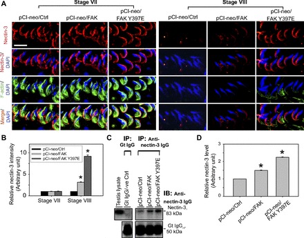
FAK Y397E mutant-induced defects in spermiation in the rat testis are mediated by retention of nectin-3 at the apical ES. A: changes in the stage-specific and spatiotemporal expression of nectin-3 (red) and F-actin (green) in the seminiferous epithelium following overexpression of pCI-neo/FAK Y397E and pCI-neo/FAK vs. control (pCI-neo empty vector) testes. In control rat testes, nectin-3, an adhesion integral membrane protein at the apical ES expressed exclusively by elongating/elongated spermatids, was prominently expressed at the apical ES in stage VII tubules, most predominantly at the convex side of the entire spermatid head and few staining at the concave side. However, the expression of this adhesion protein was considerably downregulated and virtually undetectable at the apical ES in stage VIII tubules, correlating with the onset of spermiation. However, in rat testes with overexpression of FAK and FAK Y397E phosphomimetic mutant, while changes in nectin-3 in stage VII tubules in both groups vs. the control empty vector group were negligible, a disruption on the stage-specific and spatiotemporal expression of nectin-3 in FAK-overexpressed rat testes was detected, and this effect was considerably more pronounced in the FAK Y397E phosphomimetic mutant rat testes. Nectin-3 remained highly expressed in late stage VIII tubules, colocalized partially with F-actin in pCI-neo/FAK Y397E testes. Nuclei were visualized with DAPI (blue). Scale bar = 15 μm, which applies to other micrographs. B: this histogram summarizes results shown in A illustrating the intensity of nectin-3 signals (red) at the apical ES in step 19 spermatids. Each bar is a mean ± SD of 300 randomly selected spermatids from 4 rat testes (i.e., ∼80 spermatids/rat testis) in which the level of nectin-3 in pCI-neo/Ctrl in stage VII tubules was arbitrarily set at 1. pCI-neo/FAK and pCI-neo/FAK Y397E were compared with the control group in either stage VII or VIII tubules. *P < 0.05. C: to further validate data shown in A and B and due to the limited protein loading capacity of SDS-polyacrylamide gel (∼200 μg protein/lane), nectin-3 restricted to step 18–19 spermatids in the rat testis was pulled down by Co-IP with ∼800 μg protein/sample using an anti-nectin-3 IgG (Table 1), and the immunocomplexes were used for immunoblotting (IB). Data were analyzed and shown in D. *P < 0.05.