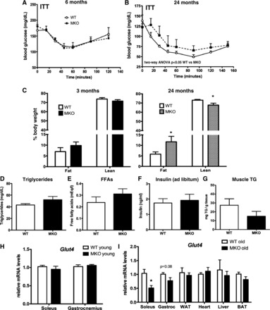
Fig. 5.

Aging promoted insulin resistance, increased adipose tissue mass, and lowered respiratory exchange ratio (RER) in MKO mice. A and B: insulin tolerance tests (ITT) in 6- (A) and 24-mo-old (B) WT and MKO mice (n = 5). C: body composition of mice by NMR (n = 6–8). D–G: triglyceride (D), free fatty acid (FFA; E), insulin (F), and intramuscular triglyceride (TG) content (G) measured by colorimetric assay using serum from fed WT and MKO mice at 24 mo of age (n = 5–9). G and I: Glut4 mRNA expression in tissues from young (3 mo, n = 8–9; H) and aged (24 mo, n = 5; I) WT vs. MKO mice. Data points represent means ± SE. *P < 0.05 WT vs. MKO. Gene expression is normalized to mean expression of corresponding tissue in age-matched WT mice. WAT, white adipose tissue; BAT, brown adipose tissue.
