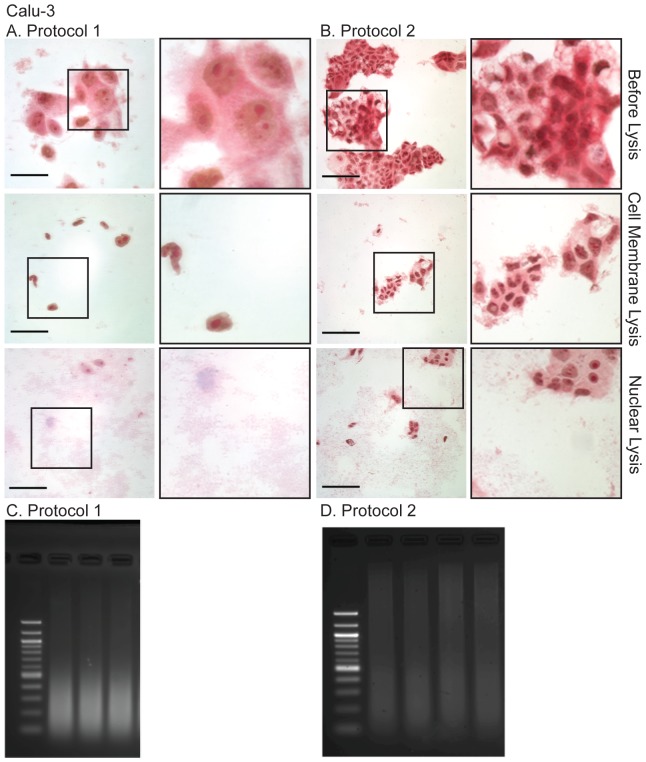
Figure 2. Light microscopy of the Methyl Green-Pyronin stained human airway epithelial cell line, Calu-3 during the modified (A, Protocol 1) and unmodified (B, Protocol 2) and resulting sonicated chromatin (C, D), separated on 2% agarose gels post-stained with 0.01% ethidium bromide.
Note, the sheets of cells have not lysed during protocol 2 and the resulting sheared DNA is of poor quality for ChIP. Methods as described in the text. Scale bar = 50 µm for A (Protocol 1) and 100 µm for B (Protocol 2).

