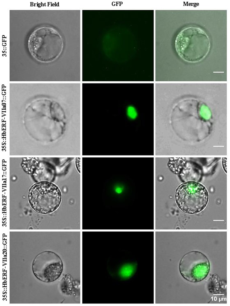
Figure 9. Subcellular localization of HbERF-VIIs.
Tree HbERF-VII GFP fusion proteins (HbERF-VIIa7, HbERF-VIIa17 and HbERF-VIIa20) were transiently expressed in protoplasts from BY-2 tobacco cells under the control of the 35S promoter. Subcellular localization was analysed by confocal laser scanning microscopy. The merged pictures of the green fluorescence channel (middle panels) and the corresponding bright field (left panels) are shown (right panels). Control cells expressing fluorescence absence are shown in the top panel. The scale bar indicates 10 µm.

