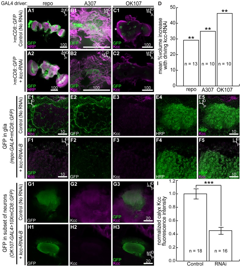
Figure 3. kcc knockdown leads to brain volume increases in 24–48 h-old adults.
Shown are representative brain enlargements via kcc RNAi in 0.5 µm confocal slices, (A2), (B2), (C2), with respect to controls, (A1), (B1), (C1), for the repo, A307, and OK107 GAL4 drivers. *: optic lobes of test or control brains were often severed to distinguish genotypes stained in same solutions. (D) Quantification of mean %volume increases for test genotype brains compared to their respective controls. Expressing kcc RNAi in glia or neurons causes whole-brain swelling. High magnification of glia with membrane-bound GFP in the wild-type mushroom body region (E2) wraps and interlaces neuronal somata and the calyx neuropile (E4). Dorsal Kcc in this same region (E3) colocalizes with cortex and surface glia (E1) and with glomeruli of the calyx (E5). Glia expressing membrane-bound GFP and kcc-RNAi-B in similar mushroom body regions (F2) are largely absent; defined cortex glia, glia wrapping the calyx, and stereotyped surface glia are few, and thus, Kcc colocalization is reduced (F1). Kcc in this region (F3) mostly colocalizes with the neuronal calyx (F4) neuropile (F5). Neuronal expression of membrane-bound GFP in the mushroom body (G1) confirms Kcc localization (G2) in the somata and calyx neuropile (G3). (H1) kcc RNAi and membrane-bound GFP expression in the mushroom body caused a significant reduction of Kcc (H2) in the calyx (H3). Moreover, brain surface Kcc is further from the calyx in this loss-of-function genotype, as seen in other genotypes with neuronal kcc RNAi expression (data not shown). (I) Quantification of Kcc knockdown in the mushroom body calyx due to kcc RNAi expression. Significance for Student's t-tests is: ** = p<0.01; *** = p<0.001. Scale bars are in microns.

