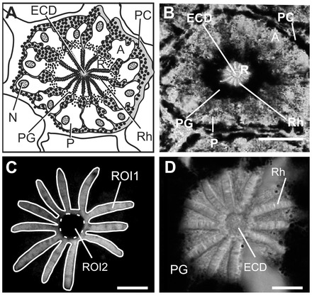
Fig. 2.

Structure of Limulus lateral compound eye (LE) ommatidia seen in cross-section. (A) Schematic of a cross-section through one LE ommatidium with 12 photoreceptor cell bodies (P). The following are labeled: A, arhabdomeral segment; ECD, eccentric cell dendrite; N, nucleus; PC, pigment cell; PG, pigment granules in photoreceptors; R, rhabdomeral segment; Rh, rhabdom. (B) Low-power brightfield image of a cross-section of an ommatidium from a LE fixed during the day. Pigment granules are distributed throughout the cytoplasm of the A segment and concentrated at the junction between the R and A segments. Scale bar, 50 μm. (C) Confocal image of Gqα-immunoreactivity in the R segment and proximal A segment of an ommatidium for a LE fixed during the day. Shown are the regions of interest (ROIs) drawn to quantify average immunoreactive intensities over rhabdoms. Total intensity of ROI1 minus ROI2 was divided by total area of ROI1 minus ROI2 to calculate the average intensity over rhabdoms per μm2. Scale bar, 10 μm. (D) Brightfield image of the section shown in C showing the rays of the rhabdom in the R segment surrounded by a dense accumulation of PGs at the junction between the R and A segments of photoreceptors. Scale bar, 10 μm.
