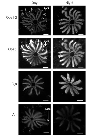
Fig. 3.

Immunoreactive intensities of Ops1-2, Gqα and Arr over rhabdomeres changed significantly from day to night while that of Ops5 did not. Representative images of single confocal optical sections showing Ops1-2-, Ops5-, Gq α- and Arr-ir in the R segment and proximal A segment of LEs (Fig. 2) fixed midday, ~30 min after peak illumination, and during the night, between 4 and 6 h after sunset. Images show the extremes of day-to-night structural changes in rhabdoms as viewed in cross-section. Daytime and nighttime images of each antigen were taken from frozen sections that were immunostained at the same time and imaged during a single confocal session with identical confocal settings. Day and night images of each antigen were intensified in Photoshop to exactly the same extent and then assembled in CorelDraw. LDS, immunoreactive membranes shed from rhabdomeres during the day by light-driven shedding, a clathrin-mediated endocytosis involving Arr as an adaptor protein (Sacunas et al., 2002). Scale bars, 10 μm.
