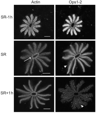
Fig. 6.

Rhabdom structure and actin- and Ops1-2-ir at rhabdoms change during dawn. Confocal images of cross-sections of LE rhabdoms double-labeled for actin-ir and Ops1-2-ir. LEs were fixed at SR-1 h, SR and SR+1 h. Sections were immunostained at the same time and images were collected in a single confocal session using identical settings. Single optical sections are shown. Images were intensified in Photoshop and assembled in CorelDraw. Images of actin-ir were intensified together and images of Ops1-2-ir were intensified together. Arrow indicates the central collar of the rhabdom. Arrowheads indicate Ops-1-2-containing debris shed by transient rhabdom shedding. Scale bars, 10 μm.
