
Fig. 4.
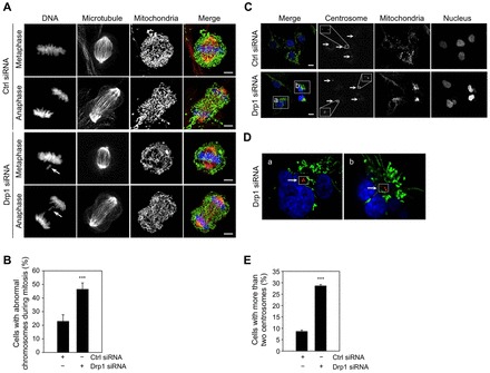
Loss of Drp1 induces chromosomal instability and centrosome overamplification. (A,B) Loss of Drp1 induces chromosome abnormalities in mitosis. (A) Mitotic chromosomes were visualized in the control cells and Drp1-deficient cells stably expressing pAcGFP1-Mito by DAPI staining. Microtubules were visualized by staining cells with Alex Fluor 555-conjugated anti-β-tubulin antibody. The images show representative cells in metaphase and anaphase. Arrows indicate lagging chromosomes. The bars indicate 5 µm. (B) The percentage of mitotic control cells and Drp1-deficient cells with abnormal chromosomes was determined by counting at least 30 mitotic cells from three independent slides. These data represent the mean ± the standard deviation (s.d.). *** P<0.005. (C-E) Loss of Drp1 induces centrosome overamplification. (C) Centrosomes in control and Drp1-deficient cells stably expressing pAcGFP1-Mito were visualized by staining cells with anti-γ-tubulin antibody, followed by secondary Alex Fluor 594 goat anti-mouse antibody. The nuclei were visualized by DAPI staining. Arrows indicate the centrosomes. The bars indicate 10 µm. (D) Enlarged images of box a and box b in panel C of a single focal plane from Drp1 knockdown cells. (E) The percentage of control and Drp1-deficient cells with more than two centrosomes was determined by counting at least 100 cells from three independent slides. These data represent the mean ± the standard deviation (s.d.). ***P<0.005.