
Fig. 3.
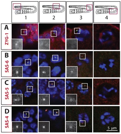
Distribution of centriolar proteins. Young adult hermaphrodite gonads stained for the indicated centriolar proteins (red in the merged images and shown alone in magnified insets), IFA-1 (green) and DNA (blue). Insets are magnified twofold. Schematic representations above the panels indicate positions of regions 1-4 in the gonad. The four panels of each row do not necessarily come from the same gonad. Note that two foci are visible next to the GCN highlighted in C, region 1 and B, region 2.