
Fig. 6.
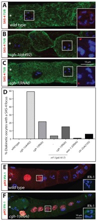
CGH-1 promotes timely centriole elimination. (A-C) Region 4 from young adult hermaphrodites of the indicated genotypes stained for α-tubulin (green), SAS-4 (red in the merged images and shown with DNA in the insets) and DNA (blue). Note that the cgh-1(ok492) diakinesis GC highlighted in B is clearly cellularized. Note also that the inset for the DNA in these panels is a projection to visualize the DNA, whereas the low magnification image represents only one of the z-sections. The most mature GC/oocyte is to the left and marked by an asterisk. (D) Percentage of diakinesis GCs with SAS-4 foci. n and P-values (compared with wild type; Fisher’s exact test): wild type, n=79; cgh-1(ok492), n=22, P<0.0001; cgh-1(RNAi), n=93, P<0.0001; rrf-1(pk1417), n=27, P=0.2547; rrf-1(pk1417) cgh-1(RNAi), n=33, P=0.0018 and P=0.6120 compared with cgh-1(RNAi); wee-1.3(RNAi), n=100, P=1; cki-2(ok2105), n=67, P=0.042. (E,F) Wild-type (E) and cgh-1(RNAi) (F) gonads of young adult hermaphrodites stained for phospho-H3 (red), IFA-1 (green in the merged images and shown with DNA in insets) and DNA (blue). Note that in ∼16% of cgh-1(RNAi) phospho-H3-positive nuclei an IFA-1 focus was observed (n=67) whereas no such cases were observed in the wild type (n=35). Insets are magnified twofold.