
Fig. 7.
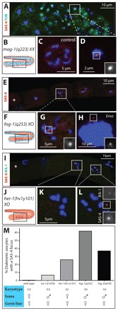
The karyotype contributes to centriole elimination. (A,C,D) Region 3/4 from a mog-1(q233)XX animal (A,D) stained for α-tubulin (green), SAS-4 (red) and DNA (blue). D shows a high magnification view of the area denoted in A, with SAS-4 signal highlighted. A heterozygous diakinetic hermaphrodite oocyte is shown in panel C for comparison. (B) Schematic representation of a mog-1(q233)XX gonad in which hermaphroditic somatic cells (red) encase male GCs (blue). (E,G,H) Region 4 from a fog-1(q253)X0 animal (E,G,H) stained for SAS-4 (red) and DNA (blue). G shows a high magnification view of the area denoted in E, with SAS-4 signal highlighted. The most mature GC/oocyte is to the left and marked by an asterisk. (F) Schematic representation of a fog-1(q253)X0 gonad, in which male somatic cells (blue) encase female GCs (red). (H) Emo oocyte. The number of SAS-4 foci in fog-1(q253)X0 animals ranged from zero to eight, both in diakinesis and Emo oocytes, with the majority having no (32% for diakinesis, 72% for Emo) or one SAS-4 focus (31% for diakinesis, 9% for Emo). We interpret the occurrence of oocytes with more than two SAS-4 foci as centrioles breaking apart or as supernumerary centriole formation during endoreduplicating cycles. (I,K,L) Region 4 from a her-1(hv1y101)X0 animal stained for SAS-4 (red), IFA-1 (green) and DNA (blue). K and L show high magnification views of the areas denoted in I. The most mature GC/oocyte is to the left and marked by an asterisk. (J) Schematic representation of a her-1(hv1y101)X0 gonad in which female X0 GCs (red) are contained in a female gonad (red). (M) Karyotype, sex of the soma and the germ line, as well as percentage of diakinesis GCs with SAS-4 foci. n and P-values (compared with wild type; Fisher’s exact test): wild type, n=79; tra-1(e1076), n=47, P=0.050; fog-1(q253), n=73, P<0.0001; fog-3(q470), n=38, P<0.0001; her-1(hv1y101), n=163, P<0.0001. Insets are magnified twofold.