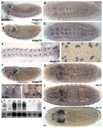
Fig. 2.

Drosophila CORL embryonic expression is restricted to CNS cells. (A) Lateral stage 13: in the brain, CORL is seen within the ventral region of the tritocerebrum (right red arrowhead) and the deutocerebrum (left red arrowhead), as well as in a central region of the ocular protocerebrum (white arrowhead). (B) Ventral 13: in the VC, one or two CORL cells per segment are detected. (C) Lateral 14: in the brain, more CORL-positive cells are present within the ventral region of the tritocerebrum (right red arrowhead), the deutocerebrum (left red arrowhead) and the ocular protocerebrum (white arrowhead). A single CORL cell dorsal to the large group of cells in the ocular protocerebrum has appeared (green arrowhead). Single cells at the dorsal/anterior tip (black arrowhead) and the ventral/medial region of the pharynx (blue arrowhead) are now visible. (D) Ventral 14: in the VC, there are groups of two or three CORL cells per segment. (E,E′) Ventral 15: in a filleted VC, sets of three CORL cells per segment (white arrowheads) are present, although groups with four or five cells are present in posterior segments. (F) Lateral 16: in the brain, CORL cells in the ocular protocerebrum now form a star-shaped pattern with the largest group in the center (filled green arrowhead) and four smaller groups located dorsal, dorsal/posterior (white arrowhead), posterior and ventral to the central group. In the anterior/dorsal (black arrowhead) and ventral/medial regions of the pharynx (blue arrowhead), as well as in the ventral region of the trito- and the deutocerebrum, CORL has expanded and the latter two have merged into a continuous row of cells (red arrowhead). The dorsal-most CORL cell has migrated to just below the dorsal surface (green arrowhead). (G) Ventral 16: in the VC, CORL persists in three to five cells per segment and new expression is observed in a single pair of cells adjacent to the midline in A3 (white arrowhead). (H-K) Dorsal views of the brain with anterior upwards in H,I and towards the left in J,K. Arrow colors indicating CORL brain expression are consistent between lateral and dorsal views. (H) Stage 13: CORL is seen in two groups of ocular protocerebral cells (white arrowheads). (I) Stage 15: in the brain, the number of CORL cells in the ocular protocerebral (white arrowheads) and medial regions (green arrowhead) increases and expression in two pairs of cells anterior to the brain is apparent (blue arrowheads). (J) Stage 16: the number of CORL cells in the ocular protocerebral region appears stable, the medial CORL cells now appear associated with the esophagus (filled green arrowhead) and a new set of paired cells at the anterior are detected (black arrowheads). (K) Stage 15: toy is visible in MB neurons in the ocular protocerebrum (white arrowheads). (L) CORL hybridized to mRNA from adult females, adult males, 0- to 12- and 12- to 24-hour-old embryos, first, second and third instar larvae, and pupae. A single transcript of 2.6 kb that corresponds in size to the CORL 5′RACE extended cDNA is detected in all lanes except L2 and L3. The blot was stripped and rehybridized with Rp49 (ubiquitously expressed) showing that that variation in CORL expression intensity is probably due to variation in RNA loading.
