
Fig. 4.
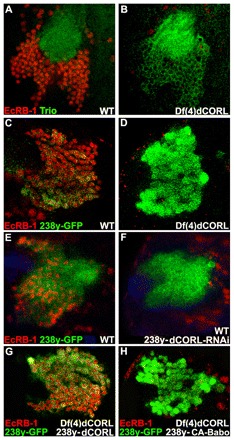
EcR-B1 MB expression is absent in Df(4)dCORL larval brains. (A,B) Ventral/posterior slice of MB cells near the calyx stained for Trio (green; membranes of all MB larval neurons but not MB neuroblasts or GMC) and EcR-B1 (red). The calyx is composed of bundled MB dendrites (green circle). In wild type, Trio and EcR-B1 (nuclear and thus not present in the calyx) are co-expressed in MB neurons. In Df(4)dCORL, EcR-B1 is absent from MB neurons (with one exception) but present in nearby non-MB neurons. (C,D) Anterior/dorsal slice of MB cells with 238y.Gal4 driving GFP (green; cytoplasmic in all MB neurons) and stained for EcR-B1 (red). In wild type, 238y-GFP and EcR-B1 are co-expressed in MB neurons but EcR-B1 is absent in Df(4)dCORL MB neurons. (E,F) Anterior/dorsal slice of wild-type MB with 238y.Gal4 driving GFP or GFP+dCORL-RNAi stained for Tll (blue; MB neuroblasts and GMC but not MB neurons), GFP (green) and EcR-B1 (red). CORL-RNAi eliminates EcR-B1 from MB neurons but not from cells outside the MB. (G) In Df(4)dCORL MB, expression of UAS.dCORL by 238y.Gal4 at 18°C rescues EcR-B1 (red) expression cell-autonomously (GFP; green). (H) Anterior/dorsal slice of Df(4)dCORL MB cells with 238y.Gal4 driving CA-Babo+GFP (green) that are stained for EcR-B1 (red). Note that loss of CORL prevents CA-Babo from activating EcR-B1.