
Fig. 4.
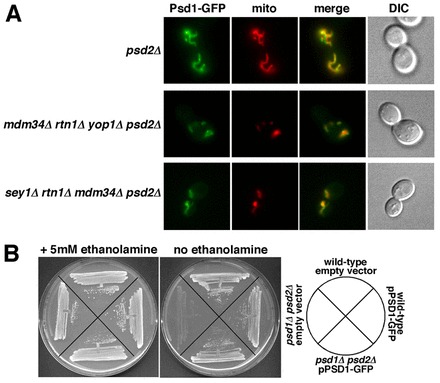
The localization of Psd1–GFP is not altered in cells lacking ER-shaping proteins and Mdm34p. (A) Cells with the indicated genotypes, expressing Psd1–GFP and Mito-Red were visualized live. (B) The Psd1–GFP fusion is functional. Strains with the indicated genotypes were plated on SC medium with or without ethanolamine. Representative examples of three independent experiments are shown.