Fig. 3.

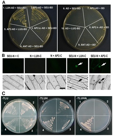
AP2 interacts with SEU but not LUG. (A) A yeast two-hybrid assay between prey (AD) and bait (BD) pairs. LEUNIG HOMOLOG (LUH)-AD against SEU-BD served as a positive control (Sitaraman et al., 2008). SEU-BD contains a truncated SEU (residues 1-563) with its C-terminal domain removed to avoid self-activation (Sridhar et al., 2006). The plate on the right shows various negative controls. The selection medium was −Trp, −Leu, −His, −Ade (−TLHA), plus 3 mM 3-amino-1,2,4-triazole. (B) A BiFC assay showing interactions in planta. Fluorescent (top row) and bright-field (bottom row) images of onion epidermal peels bombarded with BiFC plasmids. N and C represent the N- and C-terminal fragments of YFP, respectively. White arrows indicate fluorescent nuclei and black arrows indicate the same nuclei in bright field. SEU-N and LUH-C, previously shown to interact via BiFC (Hollender and Liu, 2010), serve as the positive control. Scale bar: 100μm. (C) AP2-AD and LUG-BD were tested for interaction in the presence or absence of full-length SEU expressed from the p426 vector. 1, AP2-AD + p426 vector + LUG-BD; 2, AP2-AD + p426-SEU + LUG-BD; 3, AP2delta-AD + p426-SEU + LUG-BD; 4, SEU-AD + p426 + LUG-BD (positive control). The ‘−TLU’ medium, which lacks Trp, Leu and urea, selects for all three plasmids. ‘−TLUH’ selects for His3. ‘−TLUHA’ selects for His3 and Ade2.
