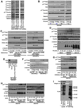
Fig. 3.

TM4SF5 activates FAK upon integrin engagement. (A) shRNA against control sequence (Con) or TM4SF5 (i.e. plain shRNA or GFP-tagged shRNA against TM4SF5) was transiently transfected to HepG2 or Huh7 cells for 48 h, before harvests of whole cell lysates for standard western blots for the indicated molecules. (B,C) SNU449 cells [SNU449 parental (P), SNU449Cp (Cp, a stable control) cells without TM4SF5 expression, but SNU449Tp (Tp, a stably pooled clone) and SNU449T16 (T16, a single cell derived stable clone) cells, B], or SNU449 cells (SNU449Cp or SNU449Tp cells) infected with adenovirus for control (Ad-TA), (HA)3-FAK WT, or myc-FERM for 24 h (C), were kept in suspension (0) or reseeded on FN for the indicated times (min), before whole-cell lysate preparation to immunoblot for the indicated molecules. (D) Stable SNU761 cells expressing mock, TM4SF5 WT, or ΔICL19 mutant were kept in suspension (0) or reseeded on FN for the indicated times. Whole-cell lysates were prepared and immunoblotted. (E) Myc-(His)6-mock or Myc-(His)6-TM4SF5 were transiently transfected for 48 h and then kept in suspension (Sus) or reseeded on FN-precoated dishes for 2 h, before preparation of whole-cell lysates for immunoprecipitation with anti-(His)6 and standard western blots for the FAK and (His)6-tag. Lysates were analyzed in parallel. (F) Cells were kept in suspension (0) or reseeded on poly-L-lysines (PL) or FN, as explained in the Materials and Methods. (G,H) SNU761 cells stably expressing mock or TM4SF5 WT were preincubated with either normal mouse IgG (Nor-IgG) or neutralizing mouse anti-human integrin β1, α5, αv antibody, before keeping in suspension (Sus) or reseeding on fibronectin for the indicated times (min), as explained in the Materials and Methods. (I) Stable SNU761 cells were harvested and immunoprecipitated with anti-FLAG antibody-precoated agarose beads, before immunoblotting for the indicated molecules in parallel with lysates. * In G and H, bands in Mock (lysate) depict nonspecific bands of anti-FLAG antibody. Data represent three different experiments.
