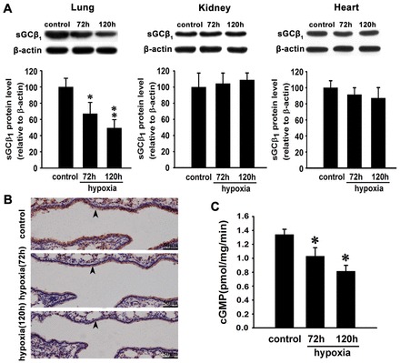
Fig. 1.

Hypoxia decreases sGCβ1 subunit expression and NO-stimulated sGC activity in mouse lung. (A) Western blot analysis and quantification of sGCβ1 subunit expression in lungs, kidneys and hearts of mice exposed to hypoxia for 72 hours or 120 hours. Expression levels are normalized to β-actin and expressed as the means ± s.e.m. (n = 4; *P<0.05, **P<0.01 versus control). (B) Immunohistochemical microscopy to assess sGCβ1 subunit expression in lungs of mice exposed to normoxia and hypoxia for 72 hours or 120 hours. Arrowheads indicate the sGCβ1-positive signals in the vascular wall. (C) NO-stimulated sGC activity in mouse lungs exposed to normoxia and hypoxia. There was a significant decrease of cGMP levels in mouse lungs after 72 hours and 120 hours of hypoxia treatment compared with normoxia-treated animals. Data are expressed as means ± s.e.m. (n = 4; *P<0.05 versus control).
