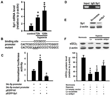
Fig. 6.

Sp1 transcriptionally regulates miR-34c-5p expression during hypoxia. (A) mRNA expression of Sp1 in mouse lungs exposed to hypoxia for 72 hours or 120 hours. Data are expressed as means ± s.e.m (n = 3; *P<0.05, **P<0.01 versus control). (B) Sp1-binding site and Sp1 expression vector. Data are expressed as means ± s.e.m. (n = 3; **P<0.05 versus empty vector group, #P<0.001 versus wild-type promoter group). (D) ChIP analysis of the miR-34c-5p promoter using pulmonary smooth muscle cells. PCR was performed to detect miR-34c-5p promoter containing a putative binding site for Sp1. (E) Western blot to confirm knockdown of Sp1 protein induced by Sp1 shRNA. (F) Western blot analysis of sGCβ1 expression in pulmonary smooth muscle cells treated with 1% O2 or Sp1 shRNA. Expression levels are normalized to β-actin and expressed as the means ± s.e.m. (n = 4; **P<0.01 versus normoxia, #P<0.05 versus Lv-scramble group).
