
Fig. 2.
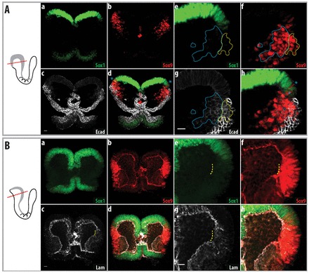
Cells delaminate from the non-neural ectoderm at ∼4 somites in mouse embryos. Delamination of cells in the non-neural ectoderm in mouse embryos. To the left are schematics of the embryos shown in the images, with the plane of section illustrated. Parts a-d of each figure show an overview, whereas e-h show a higher magnification of the neural fold. (A) Four-somite mouse embryo. Sox9 is still expressed in the non-neural ectoderm, as indicated by co-expression with E-cadherin. Sox9-positive cells that have delaminated retain their expression of E-cadherin (at lower levels, blue dotted line). Sox9-expressing cells that co-express low levels of Sox1 are indicated by blue asterisks. Sox9-expressing cells in the non-neural ectoderm are indicated by the yellow dotted line. (B) Three-somite mouse embryo, before cell delamination. Breakage of the basement membrane below the non-neural ectoderm is indicated by the yellow dotted line. Sox9-expressing cells in this region do not express Sox1. Lam, laminin α1. Scale bars: 20 μm.