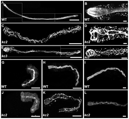
Fig. 1.

EMS mutagenesis induces mislocalization of IFB-2::CFP. (A-L) The fluorescence images show the IFB-2::CFP reporter distribution in WT background (strain BJ52; A,B,G-I) and kc2 (C,D,J-L) and kc3 (E,F) mutant worms of strains BJ133 and BJ134, respectively. (A-F) Adult worms; (G,H,J,K) embryos; and (I,L) L1 larvae. In WT adults, IFB-2::CFP localizes to the subapical perilumenal cytoskeletal network of intestinal cells (A). By contrast, in kc2 mutants IFB-2::CFP forms multiple cytoplasmic aggregates and is almost completely absent from the apical part of the cell except for a characteristic rope ladder-type pattern (C). B and D show magnifications of the anterior parts of WT and kc2 mutant adults, respectively. Young kc3 adults (<3 days) develop a less severe phenotype than kc2 with fewer cytoplasmic aggregates and only some small gaps in the subapical IFB-2::CFP network (E). In older adult worms (>3 days), however, the phenotype becomes more severe, as the gaps become bigger and cytoplasmic aggregates increase in size and number (F). In WT embryos, IFB-2 localizes to the apical membrane domain in mid-morphogenesis (G). A diffuse cytoplasmic staining can be observed which becomes weaker during further development (2-fold stage, H) and is almost undetectable in L1 larvae (I). In kc2 mutants, IFB-2 is never properly localized. During mid-morphogenesis (J) and in a 2.5-fold elongated embryo (K) only a punctate IFB-2 pattern is seen. In the L1 larvae, IFB-2 localizes in a rope ladder pattern at the cell apex and in cytoplasmic aggregates (L). Scale bars: 100 μm in A,C,E; 15 μm in G,H,J,K; 10 μm in B,D,F,I,L.
