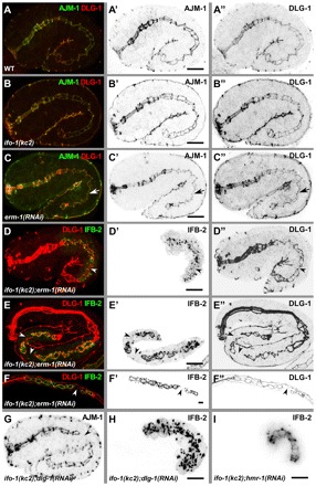
Fig. 10.

ifo-1 mutants are synthetic with erm-1 and dlg-1 mutants but not with hmr-1 mutants. (A-C″) Anti-AJM-1 (green in A-C; inverted in A′-C′) and anti-DLG-1 (red in A-C; inverted in A″-C″) immunofluorescence in 1.75-fold stages of WT (A-A″), ifo-1(kc2) (B-B″) and after RNAi against erm-1 (C-C″). Arrow indicates constriction of the future lumen. (D-F″) Fluorescence micrographs of IFB-2::CFP (green in D-F; inverted in D′-F′) and anti-DLG-1 signal (red, D-F; inverted D″-F″) in kc2 background of 1.75-fold embryo (D-D″), 3-fold embryo (E-E″) and L1-larva (F-F″) treated with dsRNA against erm-1. (G,H) Immunofluorescence of AJM-1 (G) and IFB-2 (H) in 1.75-fold ifo-1(kc2);dlg-1(RNAi) embryo. Note that the apical junctional patterns are identical in WT, ifo-1(kc2) and erm-1(RNAi) but presents disruptions in the double mutants (arrowheads in D-G) as illustrated by discontinuous DLG-1 (D″-F″) and AJM-1 (G) immunofluorescence. Also note the discontinuity in IFB-2 in ifo-1(kc2); erm-1(RNAi) (D′-F′) and the complete release of IFB-2 from junctions into cytoplasmic granules in ifo-1(kc2);dlg-1(RNAi) (H). (I) IFB-2::CFP in vivo fluorescence of an embryo treated with RNAi against hmr-1 in kc2 background. Note the continuous fluorescence pattern. Scale bars: 10 μm.
