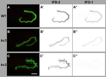
Fig. 4.

Peptide antibodies detect IFO-1 exclusively perilumenally in the intestine of WT and at reduced levels of kc3 mutants but not in the intestine of kc2 mutants. (A-C″) The images show immunofluorescence analyses (overlay at left; inverse presentation in middle and right column) of WT (A-A″), kc3 (B-B″) and kc2 (C-C″) 2-fold embryos that were stained against IFB-2 (green) and with purified anti-IFO-1 peptide antibodies (red; peptide 2, see Fig. 2B′). The micrographs that were taken under the same conditions show a decrease of intestinal IFO-1 in kc3 (B″) and a complete loss of the protein in kc2 (C). Scale bar: 10 μm.
